Genicular Neurotomy
This procedure is performed in 2 phases
1. Diagnostic Genicular Nerve Block
2. Genicular Nerve Ablation
Both portions of the procedure are performed as an outpatient under direct fluoroscopic guidance.
Diagnostic Genicular Nerve Block Procedure
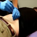
This procedure is generally well tolerated with patients often requiring only a small amount of local anesthetic. In cases where patients have particularly painful joints, light sedation or anesthesia can be provided to make patients more comfortable.
1. You will be asked to lay on your flat on your back with padding added for additional comfort.
2. The skin around your knee will be thoroughly cleaned with a sterile soap to minimize the risk of infection.
3. The target areas around the knee where the genicular nerves are suspected to be located are visualized using fluoroscopy (a real time X-ray device) and then marked on the skin.
4. Your doctor will thoroughly numb the skin and underlying tissue with local anesthetic for added comfort.
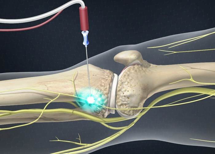
5. Thin needles will be inserted with the aid of the fluoroscopy. The target points are the confluence of the femur with the medial and lateral epicondyles and the confluence of the tibia with the medial epicondyle.
6. Once the needles are in proper position, a small amount of contrast dye will be injected to ensure the needles are in the appropriate spots.
7. At this point a small amount of local anesthetic will be injected and the procedure is complete.
After a successful blockade of the geniculate nerves, there will be a reduction in sensation in the knee as well as a slight numbness and tingling sensation around the knee.
After this portion of the procedure is completed there are 2 possible outcomes:
1. Your pain is improved or even eliminated for several hours – this indicates an obvious therapeutic value in treating the genicular nerves for your knee pain and consideration for proceeding onward to radiofreqency ablation, thus performing the genicular nerve neurotomy.
2. There is no pain relief. This will equally guide your physician’s decision making when coming up with future treatment plans.
A positive response is considered to be at least 50% pain relief for more than 24 hours following the procedure. In the event of positive response, your doctor can conclude the genicular nerves are an appropriate target in treating your knee pain and there is value in proceeding to the Radiofrequency Neurotomy portion of the treatment plan.
Genicular Nerve Ablation
Once the diagnostic portion of the procedure has been completed and a positive block has been documented, Radiofrequency Neurotomy of the Genicular Nerves is the next step. This procedure has many synonyms, all referring to the same procedure:
• Genicular RFA
• Genicular Nerve Neurotomy
• Genicular Nerve Ablation
• Genicular RF Neurotomy
This procedure is based on the theory that cutting the nerve supply to a painful structure (in this case the knee), one can alleviate pain and restore function.
As with the diagnostic nerve block described above, it is typically performed under local anesthetic but sedation can be provided where necessary. Radiodfrequency ablation of the genicular nerves is performed in a nearly identical fashion as the diagnostic nerve block. The major difference is the application of radio waves rather than local anesthetic at the conclusion of the procedure.
Special needles are used called cannulas. These cannalas are inserted in the same fashion as the needles in the diagnostic test and a just as thin. Once these cannulas are properly positioned adjacent to the genicular nerves, a small, hair-thin electrode is placed within the cannulas. To confirm the cannulas are in the correct position, your physician will “test” them by stimulating the nerves and muscles in the immediate area.
• Sensory Testing:
A painless signal will be transmitted through the electrode that will selectively stimulate sensory nerves only thus making sure there are no other nerves nearby the cannula that transmit sensation in the leg
• Motor Testing:
Completely different signal will now be transmitted that will selectively stimulate motor nerves to now make sure there are no nerves close to the cannula that are responsible for moving muscles in the leg
Once the testing is completed and your doctor has made sure the needle is safely away from any important sensory or motor nerves, some local anesthetic will be injected through the cannula to make the genicular nerves and surrounding tissue numb. Once everything is numb, radiowaves are transmitted from the tip of the cannula for 90-180 seconds. There are two different types of radiofrequency ablation that can be applied at this point:
Conventional Radiofrequency Ablation: a thermal lesion is created at 80 to 85 degrees Celsius
Cooled Radiofrequency Ablation: performed at 60 degrees Celsius, however a larger, spherical lesion is created thus decreasing the likelihood of missing the genicular nerves being targeted with the radio waves
After the radio waves are finished, the probes and cannulas are removed and 3 small dressings are applied.
The process takes less than 30 minutes to complete.